Neurosurgical procedures involving the skull often require more than completing the initial operation successfully.
In procedures such as craniotomy, decompressive craniectomy, and later cranioplasty, surgeons may also have to consider how tissue planes will heal, how easily those planes can be re-accessed during future surgery, and how scar tissue or adhesions may affect subsequent dissection.
That is where MiMedx AMNIOFIX® has become an area of interest in neurosurgery.
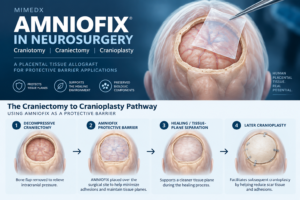
AMNIOFIX is a dehydrated human amnion/chorion membrane (DHACM) placental tissue allograft used as a protective barrier on surgical wounds. MiMedx includes craniectomies, craniotomies, and cranioplasties among the neurosurgical settings in which surgeons have used AMNIOFIX as a protective barrier.
For neurosurgeons, one particularly interesting application is the relationship between an initial decompressive craniectomy and a later cranioplasty, where preserving or establishing a manageable tissue plane may be important when the surgeon returns for reconstruction.
What Is MiMedx AMNIOFIX?
AMNIOFIX is a human placental tissue allograft manufactured by MiMedx Group, Inc.
It is composed of dehydrated human amnion and chorion membrane, commonly abbreviated DHACM.
The product is processed using MiMedx’s proprietary PURION® process, which the company describes as preserving extracellular matrix components and regulatory proteins within the placental tissue.
AMNIOFIX is also terminally sterilized for an additional level of safety and is available in multiple sheet sizes and configurations.
Within the surgical setting, MiMedx describes the primary role of AMNIOFIX as providing a protective barrier on surgical wounds.
This barrier concept is particularly relevant in neurosurgical procedures where a surgeon may later need to return through the same surgical plane.
AMNIOFIX in Neurosurgery
MiMedx currently lists several neurosurgical applications in which AMNIOFIX has been used as a protective barrier, including:
- Craniectomy
- Craniotomy
- Cranioplasty
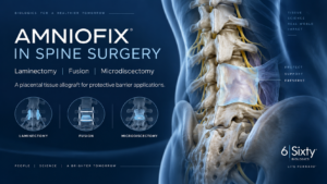
The company also lists numerous spine procedures, including:
- Laminectomy
- Fusion
- Revision surgery
- Microdiscectomy
- Endoscopic spine procedures
These applications are part of the broader interest in amniotic allografts and placental tissue allografts in neurosurgery and spine surgery.
The common theme is not that AMNIOFIX replaces the underlying surgical procedure.
Rather, the allograft is used as an adjunctive protective barrier within the surgical environment.
What Is a Craniotomy?
A craniotomy is a neurosurgical procedure in which a portion of the skull is temporarily removed to allow the surgeon access to the brain or surrounding structures.
The bone flap is generally replaced during the same operation.
Craniotomy may be performed for many reasons, including treatment of:
- Brain tumors
- Vascular abnormalities
- Hemorrhage
- Traumatic injuries
- Seizure disorders
- Other intracranial conditions
Because the surgeon must move through multiple anatomical tissue planes to access the intracranial space, postoperative healing and scar formation can influence the characteristics of those planes.
MiMedx includes craniotomy among the neurosurgical examples in which AMNIOFIX has been used as a protective barrier.
What Is a Decompressive Craniectomy?
A decompressive craniectomy differs from a craniotomy because the removed section of skull is not immediately replaced.
The procedure is often performed when dangerous intracranial pressure or brain swelling requires additional space.
Causes can include:
- Traumatic brain injury
- Intracranial hemorrhage
- Stroke-related cerebral edema
- Other conditions associated with severe intracranial pressure
After the acute problem has stabilized and swelling has resolved, the patient may later undergo a second procedure known as cranioplasty to reconstruct the skull defect.
This creates a unique surgical situation:
The surgeon knows from the beginning that there may be a second operation through the same general tissue planes.
That makes the quality and accessibility of those tissue planes especially relevant.
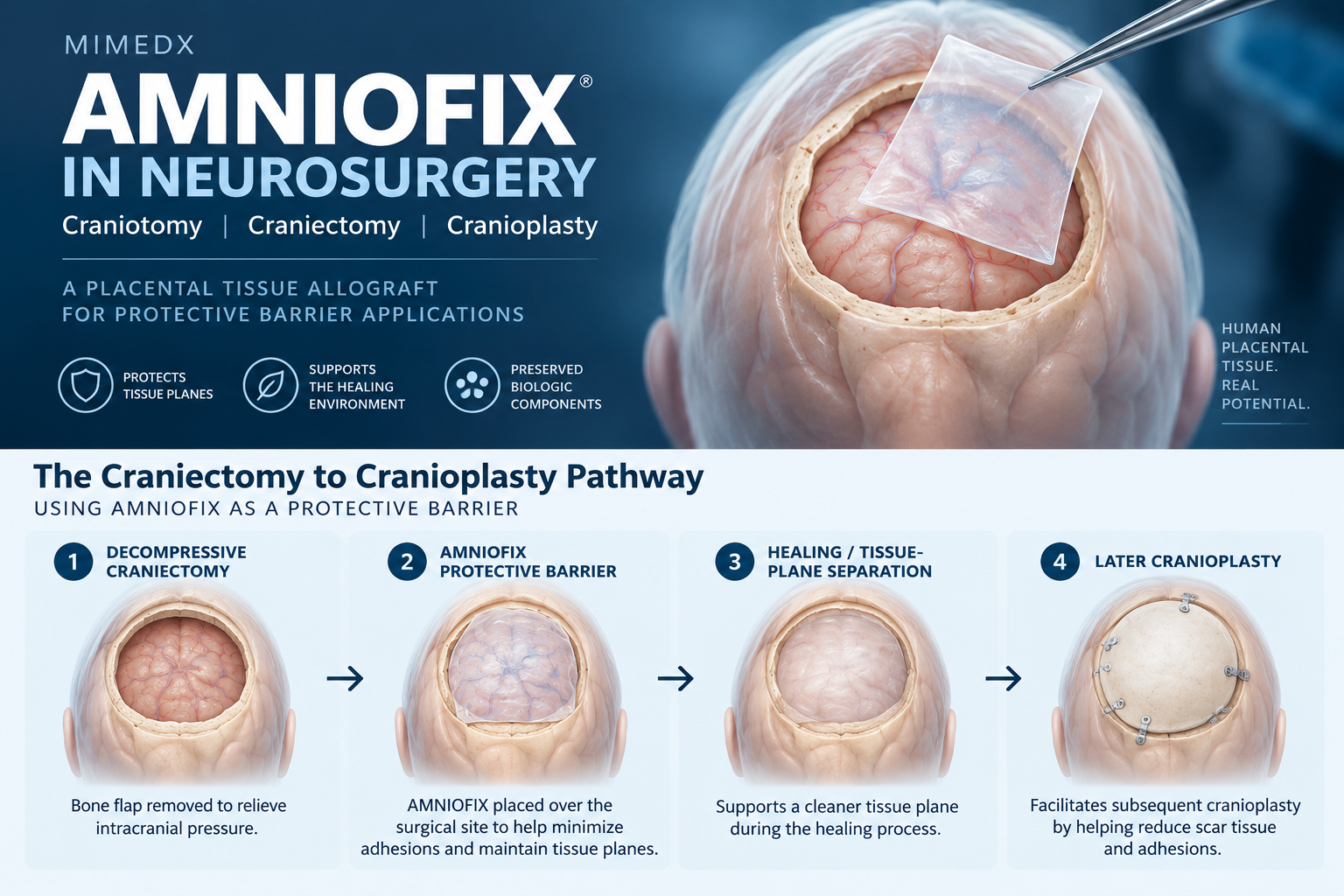
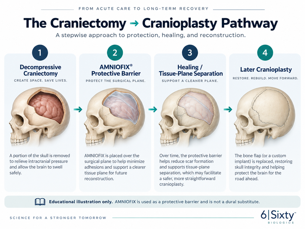
The Craniectomy-to-Cranioplasty Pathway
Place the “Craniectomy to Cranioplasty Pathway” infographic here.
Suggested caption:
Educational illustration of the decompressive craniectomy-to-cranioplasty pathway and the use of MiMedx AMNIOFIX as a protective barrier within the surgical plane. AMNIOFIX is not intended to repair dura mater and is not a dural substitute.
A simplified pathway looks like this:
1. Decompressive craniectomy
A portion of the skull is removed to provide additional intracranial space.
2. Protective-barrier placement
AMNIOFIX may be placed within the surgical field as a protective barrier according to surgeon judgment, current product labeling, and applicable Instructions for Use.
3. Healing interval
The patient heals while the skull defect remains open or protected pending reconstruction.
4. Subsequent cranioplasty
The surgeon later re-enters the region to replace the original bone flap or place a patient-specific cranial implant.
It is during that later re-entry that the condition of the tissue planes can become clinically important.
Why Tissue-Plane Separation Matters in Cranioplasty
Cranioplasty may occur weeks or months after the initial decompressive craniectomy.
During that interval, scar tissue and adhesions can develop within the operative field.
The dura may become adherent to overlying tissues, potentially making surgical re-entry more difficult.
Published neurosurgical literature has identified epidural fibrosis and dural adhesions as concerns during staged cranial procedures because difficult re-dissection can increase operative complexity.
This is one reason protective barriers have been investigated in staged cranial surgery.
The concept is straightforward:
Create or preserve a defined tissue plane during the first operation that may be easier to identify when the surgeon returns for the second operation.
Published Evidence: DHACM in Emergent Craniectomy and Later Cranioplasty
A 2023 peer-reviewed study published in the Journal of Wound Care evaluated the use of dehydrated human amnion/chorion membrane — AMNIOFIX — during emergent craniectomy procedures.
Seven patients underwent emergent craniectomy with placement of DHACM.
When those patients later returned for cranioplasty, the investigators evaluated the degree of adhesion encountered during re-exposure of the cranial defect.
The study reported:
- Negligible adhesions during defect exposure
- No intraoperative complications during the subsequent cranioplasties
- Less than three minutes devoted to exposing the defect for bone-flap placement
- Average estimated blood loss of 64.2 mL
- Average total operative time of 79.2 minutes
The study authors concluded that DHACM placement in this small patient series was associated with minimal dural adhesions during later cranioplasty.
Importantly, this was a small seven-patient observational study, not a large randomized controlled trial.
The findings are therefore interesting and clinically relevant, but they should not be interpreted as definitive evidence that every patient or neurosurgical procedure will experience the same outcome.
AMNIOFIX in Cranioplasty
A cranioplasty reconstructs a skull defect following a previous craniectomy.
Reconstruction may use:
- The patient’s original bone flap
- A patient-specific synthetic implant
- Other cranial reconstruction technologies
Before reconstruction can occur, the surgeon must safely expose the cranial defect and underlying tissues.
If extensive fibrosis or adhesions are present, this dissection may become more difficult.
MiMedx specifically highlights AMNIOFIX in the context of staged craniectomy and cranioplasty and provides surgeon case examples demonstrating the use of the graft as a protective barrier during the initial craniectomy.
The company’s educational materials emphasize the potential importance of preserving a recognizable tissue plane for later surgical re-entry.
AMNIOFIX in Craniotomy
Craniotomy is somewhat different because the bone flap is generally replaced during the same operation.
Nevertheless, MiMedx also lists craniotomy among the neurosurgical procedures in which surgeons have used AMNIOFIX as a protective barrier.
The underlying concept remains consistent:
AMNIOFIX is placed within the surgical environment as a placental tissue protective barrier, rather than as a structural cranial implant or dural replacement.
What AMNIOFIX Is — and What It Is Not
This distinction is important.
AMNIOFIX is:
- A human placental tissue allograft
- Dehydrated human amnion/chorion membrane
- A protective barrier used on surgical wounds
- Part of the broader category of amniotic allografts and surgical biologics
- Processed using MiMedx’s PURION technology
AMNIOFIX is not:
- A cranial implant
- A skull-replacement device
- A fixation system
- A dural substitute
- Intended to repair dura mater
MiMedx specifically states that AMNIOFIX is not intended to repair the dura mater and is not intended as a dura substitute.
That distinction should remain clear when discussing the product in neurosurgery.
Why Placental Tissue Is Being Explored in Neurosurgery
Human placental tissues have a distinctive extracellular matrix structure and contain naturally occurring proteins and other biological components.
Those characteristics have generated interest in placental tissue allografts across numerous surgical specialties.
In neurosurgery, much of the interest centers around their potential role as protective barriers within surgical tissue planes.
This differs from technologies whose primary purpose is structural reconstruction.
For example:
A cranial implant restores the structural defect in the skull.
AMNIOFIX serves a different purpose as a biologic protective barrier within the surgical environment.
The two technologies may therefore occupy entirely different roles within the same staged cranial procedure.
AMNIOFIX and Patient-Specific Cranioplasty
Modern cranioplasty increasingly involves patient-specific cranial implants, including materials such as PEEK, titanium, and other custom implant technologies.
These implants are designed to reconstruct the skull defect.
AMNIOFIX should not be viewed as an alternative to those implants.
Instead, the two technologies address different aspects of the surgical pathway:
Cranial implant: structural reconstruction.
AMNIOFIX: protective biologic barrier within the surgical tissue environment.
That distinction illustrates the broader evolution occurring in surgery: mechanical technologies and biologic technologies can potentially serve complementary roles rather than competing ones.
What Should Neurosurgeons Consider When Evaluating an Amniotic Allograft?
When evaluating AMNIOFIX or another placental tissue allograft in neurosurgery, surgeons and healthcare organizations may want to consider:
1. Intended use
What does the current product labeling and Instructions for Use state?
2. Tissue composition
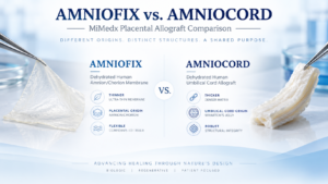
Is the graft composed of amnion, chorion, umbilical cord, or another placental tissue configuration?
3. Processing
How is the tissue processed and preserved?
4. Sterilization
Does the product undergo terminal sterilization?
5. Handling
How easily can the graft be positioned within the intended surgical field?
6. Available sizes
Is an appropriate graft size available for the anticipated surgical anatomy?
7. Published evidence
What clinical or peer-reviewed evidence exists for the specific product and application?
8. Future surgical access
In procedures where re-operation is anticipated, could preservation of a defined tissue plane be clinically relevant?
These questions are increasingly important as biologic technologies become more common in neurosurgical and spine procedures.
AMNIOFIX, Neurosurgery and the Broader Surgical Biologics Field
The use of AMNIOFIX in neurosurgery demonstrates how the field of surgical biologics is expanding beyond traditional wound-care applications.
Surgeons today have access to increasingly specialized biologic technologies designed for use across:
- Neurosurgery
- Spine surgery
- Orthopedics
- OB-GYN
- Colorectal surgery
- Urology
- Vascular surgery
- Plastic and reconstructive surgery
- Other surgical specialties
Placental tissue allografts such as AMNIOFIX are one segment of that larger biologics landscape.
For neurosurgeons, the value proposition is particularly interesting because certain cranial procedures involve planned staged surgery and future re-entry into the same operative field.
That makes tissue-plane management an especially relevant area for continued research.
Frequently Asked Questions
Is AMNIOFIX used in neurosurgery?
MiMedx lists craniectomy, craniotomy, and cranioplasty among the neurosurgical settings in which surgeons have used AMNIOFIX as a protective barrier.
Is AMNIOFIX a dural substitute?
No. MiMedx specifically states that AMNIOFIX is not intended to repair dura mater and is not intended as a dural substitute.
What is AMNIOFIX made from?
AMNIOFIX is a dehydrated human amnion/chorion membrane placental tissue allograft.
Why might AMNIOFIX be used during a decompressive craniectomy?
One area of interest is use as a protective barrier within a surgical tissue plane that may later need to be re-entered during cranioplasty.
Is there published evidence involving AMNIOFIX in craniectomy and cranioplasty?
Yes. A 2023 peer-reviewed observational study evaluated seven patients who received AMNIOFIX during emergent craniectomy and were later assessed during cranioplasty. The investigators reported minimal dural adhesions and rapid exposure of the cranial defect during re-entry.
Is AMNIOFIX the same thing as a cranioplasty implant?
No. A cranial implant reconstructs the skull defect. AMNIOFIX is a placental tissue allograft used as a protective barrier within the surgical environment.
The Growing Role of Amniotic Allografts in Neuro and Spine Surgery
The intersection of amniotic allografts, neurosurgery, and spine surgery represents an important area of growth within surgical biologics.
MiMedx currently positions AMNIOFIX and other placental tissue products across several neuro and spine applications.
As additional clinical evidence develops, surgeons and healthcare organizations will be able to better define where biologic protective barriers provide the greatest value.
For 6|Sixty Biologics, this is an important area of educational focus because neurosurgery and spine represent specialties where advanced mechanical technologies and advanced biologic technologies increasingly intersect.
About 6|Sixty Biologics
6|Sixty Biologics is an independent surgical biologics distributor focused on connecting surgeons and healthcare organizations with advanced biologic technologies across multiple surgical specialties.
6|Sixty works with MiMedx surgical biologics, including AMNIOFIX, while providing product education, hospital support, and experienced surgical representation.
Our growing educational library explores:
- AMNIOFIX
- Amniotic allografts
- Placental tissue allografts
- Surgical biologics
- Neurosurgery
- Spine surgery
- OB-GYN
- Orthopedics
- Colorectal surgery
- Urology
The goal is to help surgeons, hospitals, and industry professionals better understand where emerging biologic technologies fit within modern surgical care.
For more information on 6|Sixty, visit https://6sixty.com
References and Additional Information
MiMedx — Spine and Neurosurgery
Manufacturer information regarding AMNIOFIX and other placental allografts used as protective barriers in spine and neurosurgical settings.
MiMedx — AMNIOFIX
Official manufacturer product information regarding AMNIOFIX composition, processing, and product characteristics.
Endicott L, Ehresman J, Tettelbach W, Forsyth A, Lee BS.
Dehydrated human amnion/chorion membrane use in emergent craniectomies shows minimal dural adhesions. Journal of Wound Care. 2023;32(10):634-640.
Important Information
This article is provided for educational purposes only and is not intended to provide medical advice or direct clinical decision-making.
Product selection and use should be determined by qualified healthcare professionals after reviewing current product labeling, Instructions for Use, institutional requirements, patient-specific circumstances, and available clinical evidence.
6|Sixty Biologics is an independent surgical biologics distributor and is not MiMedx. AMNIOFIX®, PURION®, and MiMedx are trademarks or trade names of MiMedx Group, Inc.